SPECIALTY PROGRAM IN ORTHOPEDICS AND TRAUMATOLOGY
5-YEAR RESIDENTIAL TRAINING PROGRAM
STUDY PLAN AND PROGRAMS

Area I. Emergency care
This area of care is responsible for everything related to the examination, diagnosis, prognosis and emergency treatment of traumatic injuries, open or closed, of the limbs, spine, skull, brain, spinal cord and facial massif
This residency comprises the modules
- Traumatic injuries of the limbs and spine
- Traumatic injuries of the brain and spinal cord
- Maxillofacial traumatic injuries
Residency lasts 3 months and is the beginning of the specialization process. During this time, on-duty care teams complete rotations in care of adult and children with systemic multiple injuries, neurotrauma, maxillofacial traumatology, and intensive care units.
Module 1. Traumatic injuries of the limbs and spine
Objective: Faced with patients with traumatic injuries of the limbs and spine, establish a diagnosis, prognosis and emergency treatment in accordance with regulations, based on the clinical history and a physical examination, aided by complementary examinations
Assess the severity of patients and help with emergency surgeries
Skills Knowledge
Carry out the questioning, physical examination and presumptive diagnosis of traumatic limb injuries
- Collect the history of the trauma or injury through questioning (anamnesis)
- Concept of traumatized and polytraumatized (multiple systemic trauma) and their medical and socio-economic importance in times of peace and war
- Endocrine-metabolic response to trauma
Skills
- Detect skeletal discontinuity of the limbs in unconscious patients by evaluating segmental instability
- Detect pain and functional impairment of the limbs in conscious patients and differentiate it from neurovascular deficits
- Detect soft tissue discontinuity: skin, subcutaneous cellular tissue (SCT), aponeurosis and musculotendinous masses
- Detect alterations in the normal morphology of the limbs that accompany fractures and dislocations:
- Angulations of the limb axis
- Reductions
- Abnormal limb rotations
- Detect subsequent and delayed changes in volume and color of sprains, fractures and dislocations of the limbs
- Detect traumatic tendon injuries of the limbs
- Detect indirect signs of spinal injuries that are not accompanied by neurological deficit
- Pain
- Muscle spasm
- Deformity
- Functional limitation of spinal mobility
Knowledge
Concept of osteoarticular disorders
Classification of fractures and dislocations
Semiology and semiotics of the musculoskeletal system (SOMA), with regard to the limbs
Signs of abnormal mobility and interfragmentary crepitus
Concept of functional impairment and its pathogenesis
Peripheral vascular and nervous semiology and semiotics
Peripheral neurovascular impairment
Concept of soft tissue discontinuity
- Concept, pathogenesis, pathological anatomy and clinical picture of open lesions due to the detachment-rolling of the skin and Morell-Lavalle serous effusion
Concept of limb axes
Clinical signs of the cited fractures and dislocations
Concept of hematoma, its pathogenesis and evolution
Concept and characteristics of volume increase (edema-swelling) and ecchymosis in limb injuries
Concept of tendon injuries
Clinical picture of traumatic tendon injuries
Concept, pathogenesis and classification of spinal injuries
Symptoms and signs of the aforementioned spinal injuries
Skills
- Act using legal medical criteria in emergency assistance to injured patients
Make a positive diagnosis:
- Indicate and interpret the simple radiological examination of anteroposterior and lateral views of the bones and joints of the limbs and spine
- Indicate and interpret hematological, biochemical and microbiological tests
- Achieve a topographical diagnosis of osteoarticular injuries, in adults and children, of:
- Diaphyseal, metaphyseal and articular fractures of the skeletal components of the upper and lower limbs
- Dislocations and fracture-dislocations of the limbs
- Fractures, fracture-dislocations and dislocations of the thoracic and pelvic belt
- Fractures, dislocations and fracture-dislocations of the spine
- Bone injuries and open joints
Knowledge
- Measures to be taken and necessary legal medical record documentation in an injured patient, especially in stab, blunt and fire wounds:
- Preservation of evidence (weapons, clothing, projectiles)
- Documentation regarding the characteristics of the wound
Normal radiological imaging (adult and child) of the bones and joints of the limbs and spine
Radiological imaging of fractures and dislocations of the limbs and spine
Normal values and alterations in hematological, biochemical and microbiological tests
- Clinical picture and diagnosis of diaphyseal and articular fractures of the upper and lower limbs
- Clinical picture and diagnosis of limb dislocations and fractures
- Clinical picture and diagnosis of fractures, dislocations and fracture-dislocations of the thoracic and pelvic belt
- Clinical picture and diagnosis of fractures, dislocations and fracture-dislocations of the spine
- Clinical picture and diagnosis of open bone and joint injuries
Skills
- Diagnose soft tissue injuries and traumatic limb amputations
- Diagnose tendon injuries
- Syndromic diagnosis of arterial insufficiency and peripheral nerve injuries (acute and traumatic) of the limbs
- Assess the severity of the injury to the limbs or spine
Institute emergency prophylactic medical treatment for open injuries (osteoarticular or soft tissue):
- Systemic and local prophylactic antibiotic therapy
- Tetanus prophylaxis
- Analgesia
Carry out treatment using simple manual techniques:
- Perform emergency immobilization of bone injuries of the limbs and spine by:
- Sling
- Velpeau bandage
- Splints (wood, metal, plastic, etc.)
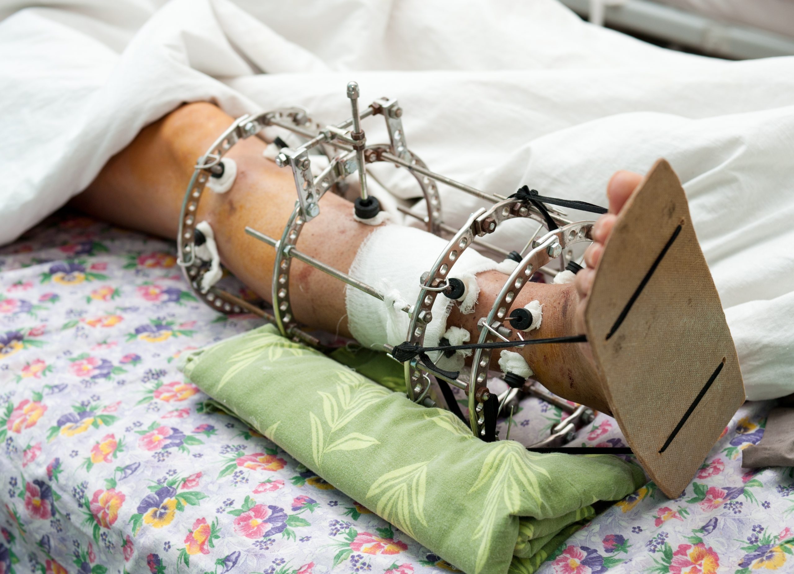
- Casts
- Full circular cast (open)
Knowledge
Clinical picture of detachment-rolling skin injuries
Clinical picture of Morrell-Lavalle serosanguinous effusion
Clinical picture and diagnosis of other soft tissue (integumentary) injuries and traumatic amputation of limbs
Clinical picture and diagnosis of tendon injuries
Clinical picture and diagnosis of acute traumatic arteriovenous insufficiency of the extremities
Clinical picture and diagnosis of acute traumatic peripheral nerve injuries
- Abbreviated scale for assessing the severity of injuries
Indications and doses of antibiotics as prophylaxis of infection
Anti-tetanus serum and tetanus toxoid: action, dosage, routes of administration and complications due to their use
Most frequently used analgesic drugs: dosage, routes of administration, complications and side effects
Technique for the indicated emergency immobilizations
Skills
- Control of external bleeding in open fractures, traumatic amputations and other bleeding injuries of the limbs by compression bandage and tourniquet
- Eliminate mechanical obstacles to arteriovenous circulation arising from previous immobilization
- Suture non-contaminated wounds involving skin, SCT and aponeurosis
Assist in emergency surgeries
Knowledge
Ischemia technique by local compression and tourniquet: indications, contraindications and complications
Technique for removing bandages (gauze, tape or plaster)
Suture technique for minor wounds (uncomplicated)
Concept and techniques of asepsis and antisepsis in the operating room
Sutures and emergency surgical instruments
Means of emergency osteosynthesis
- Principles and techniques of surgical debridement of open soft tissue injuries
Module 2.
Traumatic injuries of the brain and spinal cord
Objective: In patients with head and spinal cord injuries, establish the positive-topographic-radiological diagnosis of cranial vault fractures and the presumptive diagnosis of skull base fractures, based on clinical history, physical examination and complementary tests
Define the topographic level of the spinal cord injury and correlate it with the radiological diagnosis of spinal injury
Assess the severity of the injury and carry out emergency treatment
Carry out the interrogation, clinical examination and presumptive diagnosis of head and spinal cord injuries:
- Collect the history of the trauma or injury through questioning
- Define the different states of consciousness: normal, stupor and coma
- Detect and interpret signs of neurological deficit:
- Motor:
Hemiparesis
Paraparesis
Anisocoria
Autonomic dysreflexia, etc.
- Sensory:
Numbness
Analgesia
Hyperalgesia
Sensitivity levels
- Osteodentin, cutaneous and abnormal reflexes:
Hyporeflexia
Hyperreflexia
Babinski
- Cortical irritation:
Generalized seizures
Focal seizures
Psychomotor agitation
Concept of brain and spinal cord trauma
Neurological semiology and semiotics of head and spinal cord trauma
Concept of normal consciousness, stupor and coma
Symptoms and signs of indicated neurological deficit
Skills Knowledge
- Examine a cranial hematoma
- Examine a skull or spinal injury
- Detect the presence of clinical symptoms indicative of a skull base fracture:
- Otorrhagia
- Rhinorrhea
- Palpebral and mastoid hematomas
Make the positive diagnosis:
- Achieve a topographic-radiological diagnosis of fractures and intracranial foreign bodies, in adults and children, of:
- Linear vault fractures
- Depression fractures
- Radiopaque foreign bodies
- Achieve a (presumptive) clinical diagnosis of skull base fracture
- Correlation between the radiological bone level of vertebral injury and the clinical level of spinal cord injury
- Assess the severity of the brain and spinal cord injury
Knowledge
Clinical picture of peri cranial hematoma
Clinical picture of penetrating and non-penetrating skull-brain injuries
Clinical symptoms of indicated basilar skull fracture
Normal radiological imaging, in adults and children, of the cranial vault (anteroposterior and lateral views)
Radiological imaging of cranial vault fractures:
- Linear
- Depressed (sinking)
Imaging of foreign bodies and their topographic location
- Clinical picture of skull base fractures
Scheme of metameric segmentation and its innervation
- Abbreviated scale for assessing the severity of injuries
- Glasgow scale (or modification of it)
Skills
Carry out treatment using simple manual techniques:
- Bandage skull-brain wounds
- Immediately immobilize spinal cord injuries
- Control external bleeding in open head and spinal cord injuries using the local compression method
- Suture non-contaminated wounds involving skin, SCT and aponeurosis
Carry out emergency prophylactic medical treatment in open head or spinal cord injuries:
- Systemic prophylactic antibiotic therapy
- Tetanus prophylaxis
Knowledge
Capeline bandage technique
Immobilization technique and emergency transport in spinal cord injuries
Local compression ischemia technique: indications, contraindications and complications
Suture technique for minor wounds (uncomplicated)
Indications and doses of antibiotics as prophylaxis of infection
Anti-tetanus serum and tetanus toxoid: action, dosage, routes of administration and complications due to their use
Skills
Knowledge
Module 3.
Maxillofacial traumatic injuries
Objective: Faced with patients with maxillofacial trauma, establish the clinical and radiological diagnosis of injuries to the facial skeleton, as well as the clinical diagnosis of mechanical obstructive ventilatory failure, based on the interrogation and clinical examination
Carry out emergency treatment for these maxillofacial injuries and respiratory distress and assess their severity
Carry out the questioning, physical examination and presumptive diagnosis of maxillofacial traumatic injuries:
- Collect the history of the trauma or injury through questioning (anamnesis)
- Detect the symptoms of mechanical obstructive ventilatory failure
- Detect alterations in the normal morphology of the face
Make the positive diagnosis:
- Indicate and interpret the simple radiological examination in anteroposterior, lateral and oblique views
- Diagnose ventilatory failure caused by injuries to the facial skeleton
- Diagnose facial bone injuries
- Assess the severity of maxillofacial injuries
Concept of maxillofacial injuries
Maxillofacial semiology and semiotics
- Clinical picture of mechanical obstructive ventilatory failure
Deformities of the face determined by different maxillofacial injuries
Normal radiological imaging, in adults and children, of the facial bones
Radiological imaging of maxillofacial fractures
- Clinical picture of mechanical obstructive ventilatory failure
- Clinical picture of facial bone injuries
- Abbreviated scale for assessing the severity of injuries
Skills
Carry out emergency prophylactic medical treatment in open maxillofacial injuries:
- Systemic and local prophylactic antibiotic therapy
- Preventive treatment of tetanus
Carry out treatment using simple manual techniques:
- Place the oropharyngeal tube
- Urgently stabilize the tongue
- Perform anterior nasal packing
Knowledge
Indications and doses of antibiotics as prophylaxis of infection
Anti-tetanus serum and tetanus toxoid: action, dosage, routes of administration and complications due to their use
Most frequently used analgesic drugs: dosage, routes of administration, complications and side effects
Indications and application technique of oropharyngeal tube
Indications and different methods of emergency stabilization of the tongue
Indications and technique of anterior nasal packing
Area I. Emergency care
TEACHING STRATEGY
MODULES 1, 2 AND 3
Residency in the emergency area is completed in thirteen (26) weeks, and rotations will be made during which the resident will do on-site shifts (minimum: 24 hours every 5 days), with the noted exceptions:
- On-duty emergency team for polytraumatized (adults): six (12 weeks)
- On-duty emergency team for neurotrauma two (4) weeks
- On-duty emergency team for maxillofacial traumatology: one (2) week
- Intensive care for adults: two (4) weeks
- Intensive care for children: two (4) weeks
- On-duty emergency team for polytraumatized (children): total shifts during the residency: 7
The resident will complete only one physical shift per week in this one (except for the weeks that s/he will rotate through the intensive care units for adults and children)
The optimal sequence of rotations is the one listed. However, another sequence may be taken, as long as it does not split the time of the rotations, with the exception of the rotation with the on-duty emergency team for adult polytraumatized patients, which can be divided into two parts.
TEACHING-PATIENT CARE ACTIVITIES
- Activities in on-duty emergency teams and intensive care units
- Assistance in emergency surgical interventions (instrumentalist and third assistant)
- Institutional clinical-pathology meeting
ACADEMIC ACTIVITIES
Conferences
At least two (2) will cover the following topics:
- “Assessment of the severity and prognosis of the polytraumatized patient”
- “Injuries due to the detachment-rolling of the skin of the limbs”